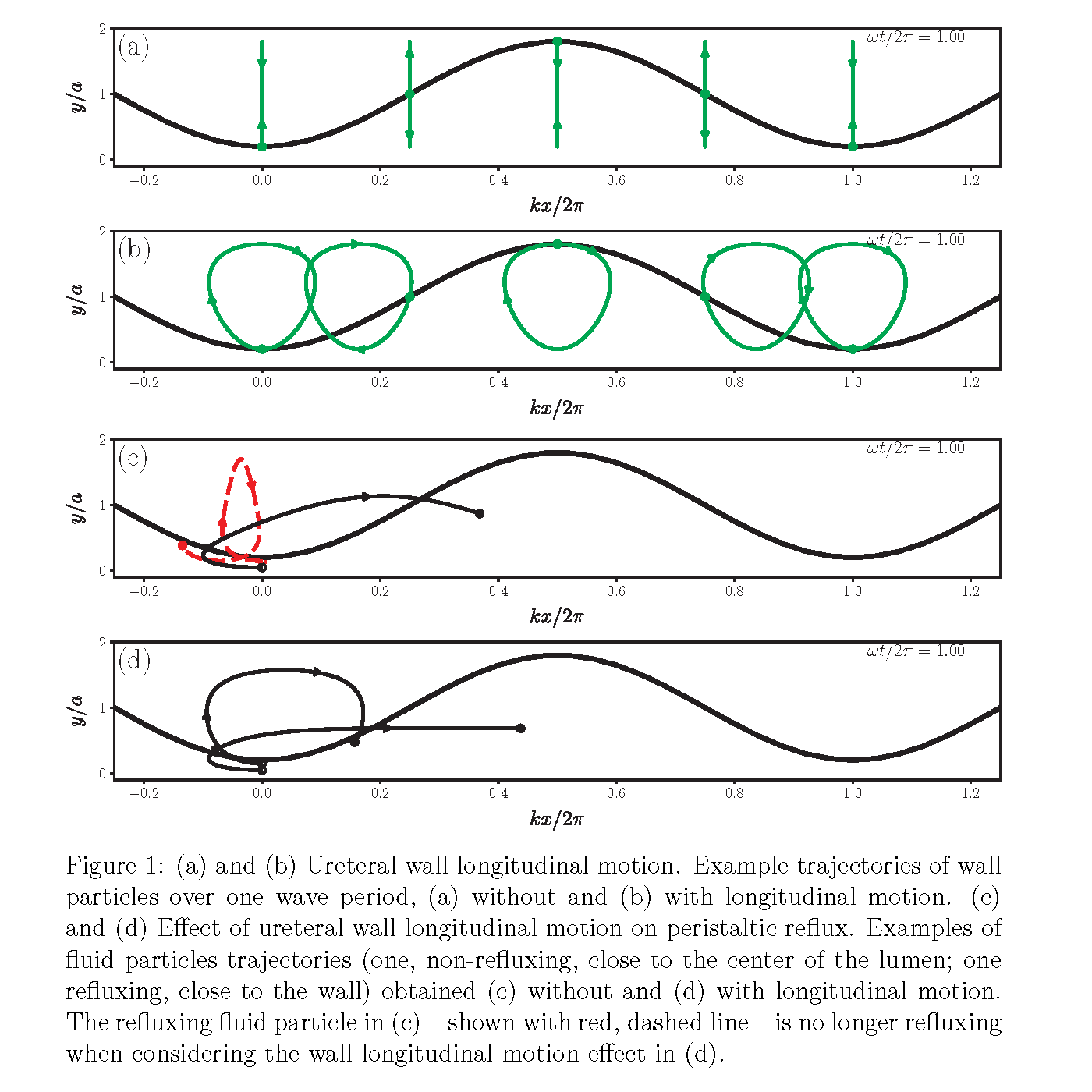
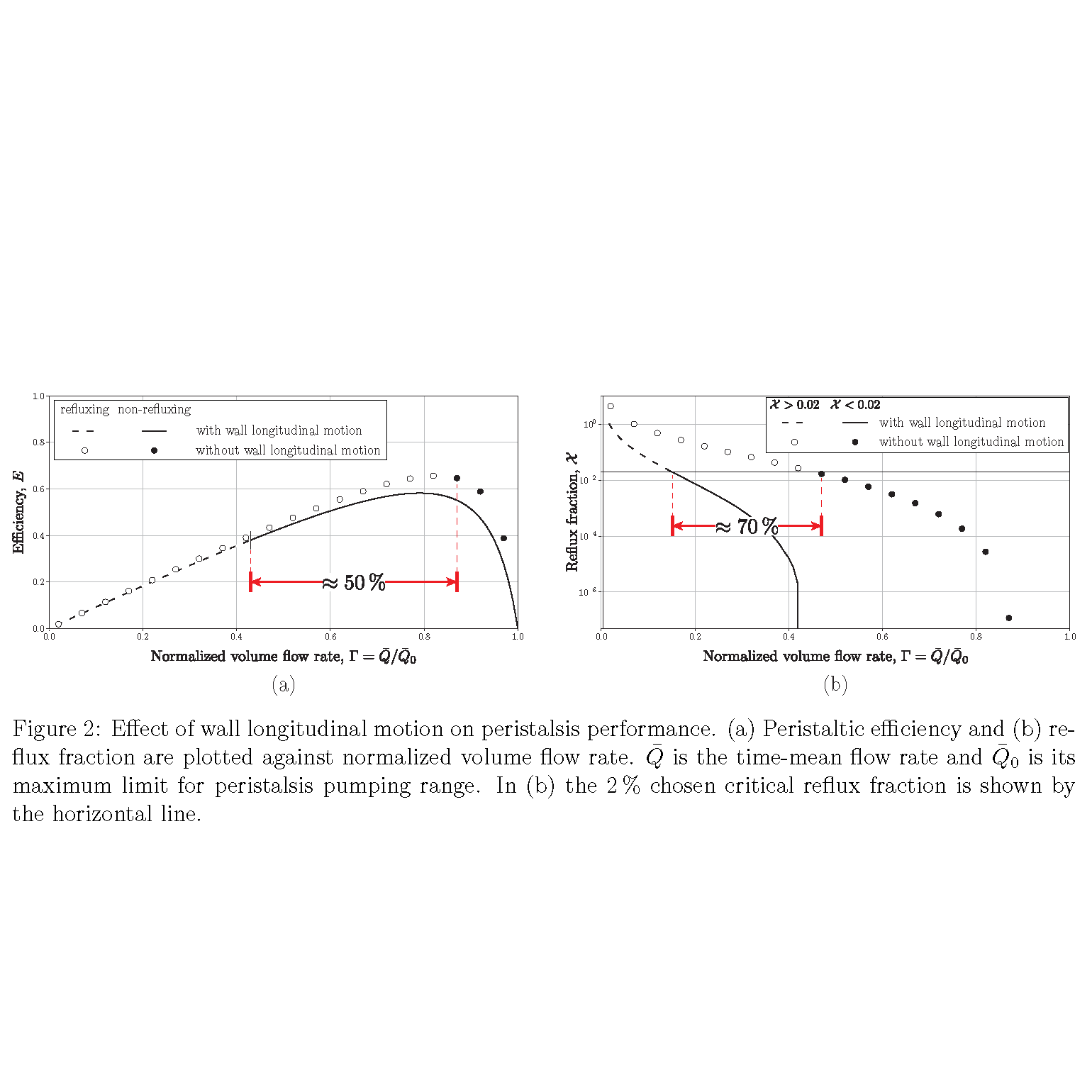
Introduction: Lack of sufficient ureteral peristalsis is theorized as one of the mechanisms contributing to ureteral dilation in vesicoureteral reflux (VUR) and non-obstructing megaureters. To help understand the phathophysiologic mechanism for these disorders, we first need to understand the dynamics of normal ureteral peristalsis, which results in near complete clearance of urine from the ureter (i.e., non-dilated). We hypothesize that the clinically observed ureteral wall longitudinal motion is crucial to prevent and/or limit peristaltic reflux (i.e., retrograde flow of urine during an episode of peristalsis). Methods: To examine this hypothesis, based on the lubrication theory in fluid mechanics, we modeled ureteral peristalsis mathematically as an infinite train of sinusoidal waves and analyzed the antegrade and retrograde flows in the ureter. In doing so, we accounted for ureteral wall longitudinal motion by imposing a periodic Gaussian longitudinal velocity on the wall, Figs. 1 (a) and (b), inspired by videos of ureteral operations. Results: Effects of wall longitudinal motion on peristaltic reflux and performance are shown in Figs. 1 (c) & (d), and Figs. 2 (a) & (b), respectively. The results are shown as an example for a typical clinical case with discharge rate of 1.62 mL/min and peristaltic amplitude of 80 % of lumen diameter. As shown, by considering wall longitudinal motion, the range of urine flow rates resulting in peristaltic reflux drops substantially. The minimum required flow rate to prevent peristaltic reflux drops by 50% when including the wall longitudinal motion effect, Fig. 2 (a). Similarly, the required flow rate to limit the reflux volume fraction below a critical value of 2% (chosen as an example, based on clinical expertise) drops by 70%, Fig. 2 (b). Conclusions: This computational model is the first to include longitudinal wall motion, which aligns with our hypothesis that it is important for minimizing peristaltic reflux. This supports the clinical observation of normal ureters having high emptying ability and helps direct further investigations to understand ureteral disorders. SOURCE OF Funding: This research has been partially supported by grants from the Michigan Institute for Clinical and Health Research (MICHR), and the Departments of Urology and Radiology at the University of Michigan.

 photo")