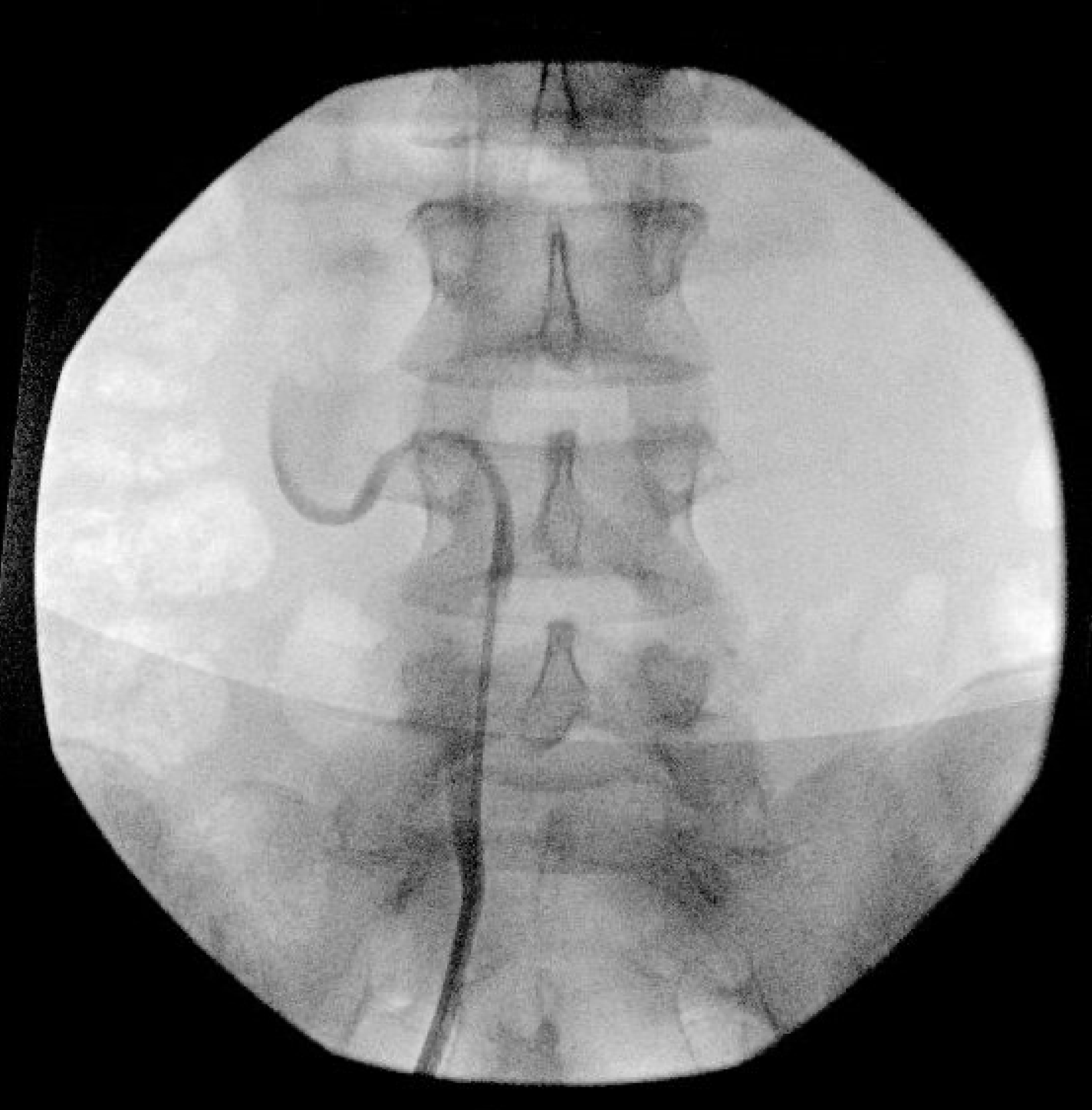
Introduction: Retrocaval ureter, also known as “circumcaval ureter” or “preureteral vena cava,” is a relatively rare anatomical variant with an incidence of 0.13% that occurs when the right ureter courses posterior to the vena cava rather than anterior. More commonly seen in males, the etiology is a developmental abnormality of the vena cava, not the ureter. Many cases are clinically silent, but surgical reconstruction is indicated if the patient has severe hydronephrosis, significant functional obstruction, or pain. This patient presented with right flank pain, severe hydronephrosis, and a characteristic "S sign" on retrograde pyelogram (Figure 1). Our objective was to describe our approach to an uncommon case, robotic ureteroureterostomy of a retrocaval ureter. Methods: The patient was placed in the left lateral decubitus position. The Da Vinci Xi system was utilized and 4 ports were placed in a linear fashion at the lateral border of the rectus 6cm apart. A 5mm AirSeal was placed between the 2nd and 3rd arm. ProGrasp Forceps are used in the 1st arm, Maryland bipolar forceps are used in the 2nd arm, and a monopolar curved scissor is used in the 4th arm. Vessel loops were placed on the proximal and distal ureter on either side of the vena cava and were pulled back and forth to allow for visualization and dissection (Figure 2). Results: Successful robotic ureteroureterostomy of a retrocaval ureter. Conclusions: Robotic ureteroureterostomy is a minimally invasive procedure to manage ureteropelvic junction obstruction caused by a retrocaval ureter. SOURCE OF Funding: None