MP09-14: Radiomic analysis to distinguish between upper tract urothelial carcinoma and renal cell carcinoma: Can we improve imaging using machine learning?
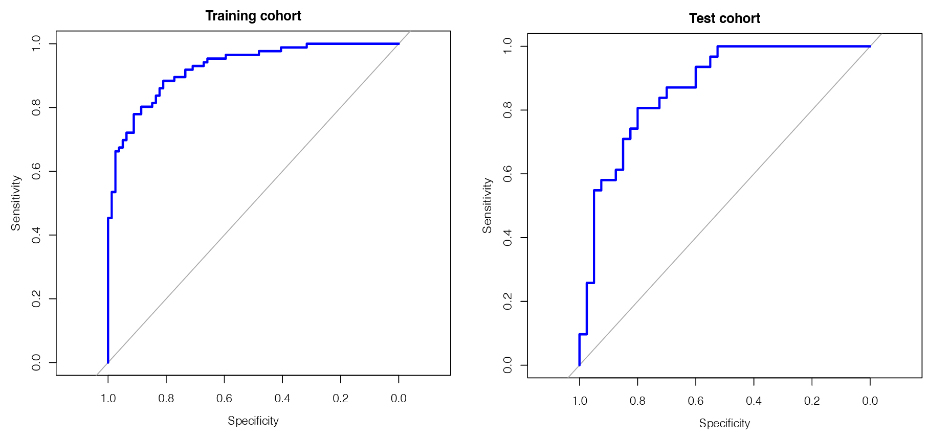
Introduction: Imaging to reliably differentiate between upper tract urothelial carcinoma (UTUC) and renal cell carcinoma (RCC) is limited in its accuracy. Radiomics uses high-throughput extraction of data from imaging series, while machine learning can be applied to identify radiomic features predictive of clinical endpoints. We aim to assess whether Radiomics and machine learning approaches might improve preoperative diagnostics to distinguish between UTUC and RCC. Methods: After IRB approval, we queried our internal database. Inclusion criteria were the diagnosis of a pathologically confirmed RCC or UTUC and available CT scans in the venous phase. Manual tumor segmentation in the axial plane was carried out by a board-certified radiologist with more than 7 years of experience following a standardized approach using imaging software mint LesionTM (Mint Medical GmbH, Heidelberg, Germany). To test for inter-reader variability (IRV), we randomly selected 30% of imaging series for which segmentation was repeated by a second board-certified radiologist. Intraclass correlation coefficient was performed to calculate IRV. Lasso regression and cross validation analyses were performed to identify predictive features to differentiate between UTUC and RCC. ROC analysis was then carried out as a goodness-of-fit test regarding predictive ability of the radiomic score. Results: 236 patients were included in this study of which 119 (50.4%) presented with RCC and 116 (49.6%) with UTUC. All patients were treated at our academic center between 2005 and 2021. 72 patients were female (30.5%) and 164 (69.5%) were male. The median age of the study cohort was 70.5 (IQR: 59.5-77) years. An IRV > 80% was determined for a total of 24 radiomic features. Using the radiomic score, a differentiation between UTUC and RCC was possible with a sensitivity of 88.4% and a specificity of 81% for the training cohort. For the test cohort, a sensitivity of 81% and a specificity of 80% was observed (Fig.1). Conclusions: Using radiomic analysis and a machine learning approach, we could reliably distinguish between RCC and UTUC. Further studies are required to determine the role of radiomic analysis and machine learning in routine imaging of unclear renal masses. SOURCE OF Funding: none