Memorial Sloan Kettering Cancer Center & Medical University of Vienna
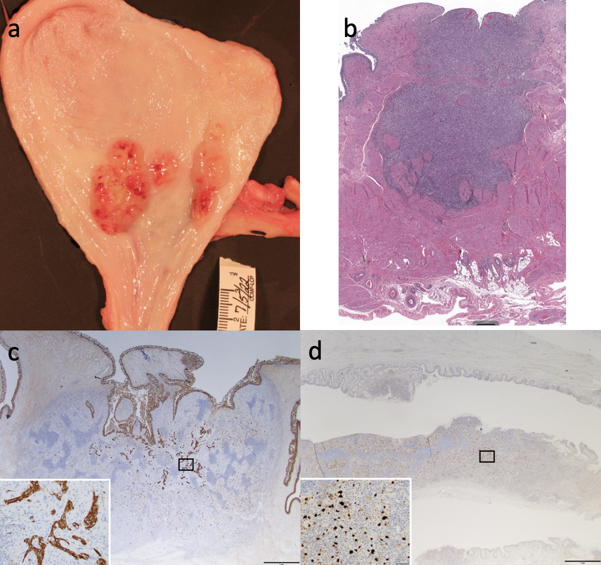
Introduction: Large animal cancer models have potential to improve and accelerate the development of new treatment modalities but few exist. Oncopigs (OPG) are particularly suitable due to their similarities to humans. We sought to develop a urothelial tumor model using the oncopig cancer model (OCM: University of Illinois Cancer Center). The OCM carries two mutations (KRASG12D and TP53R167H) which can be induced by exposure to a buffered Adeno Virus Cre solution (bAdCre). Three procedures (P1, P2, P3) were devised and investigated. Methods: Eight OPGs underwent tumor inoculation. In P1, bAdCre with plant starch (n=3) was injected submucosally at 3 distinct locations in the bladder wall. In P2 the urothelium was denuded in 3 locations followed by bAdCre instillation (n=4). In P3 bAdCre was instilled combined with N-Dodecyl- ß-D-Maltoside (DDM), an alkyl disaccharide and polar surfactant that enhances virus transfection in the bladder, with intact urothelium (n=1). Serial imaging, cystoscopies and blood draws were performed until sacrifice at 3 timepoints (days: 14, 21, 28). Tumor characteristics were assessed by necropsy and histopathological examination of the entire tumor tissue. Results: In total, 7/8 (87.5%) OPGs grew bladder tumors. Histopathological examination revealed 2 different tumor types: neoplastic tumors (NT) and inflammatory tumors (IT). Using P1, 3 OPGs developed IT (3/3). 2 OPGs in 2 locations (2/3) and 1 in 1 location (1/3). In P2, 3/4 OPGs grew tumors. 1 OPG in all 3 locations, all 3 were NTs (3/3) (Fig.1 a), 1 OPG in 2 locations (2/3), both NTs and 1 OPG in 1 location (1/3), an IT. In P3 1 NT developed (1/1). All tumors showed muscle invasive growth. P3 showed the most superficial lesion after 21 days (pT2a). In P1, 2 pigs had pT3 tumors after 21 and 28 days. No complications were observed. Based on the histological (Fig.1 b: H&E), multiple immunohistochemical findings (e.g.: Fig.1 c: high molecular cytokeratins 34Be12; Fig.1 d: P53) and the growth pattern, NT were diagnosed as urothelial cell carcinomas, sarcomatoid variant. Conclusions: This is the first description of bladder tumor development in a porcine model using the OCM. We were able to create neoplastic tumors in a short period of time. The model is reproducible and neoplastic lesions show urothelial origin with sarcomatoid transformation. SOURCE OF Funding: MSKCC Sidney Kimmel Center for Urologic Cancers

 photo")