Session: MP05: Stone Disease: Basic Research & Pathophysiology
MP05-01: MICRO COMPUTED TOMOGRAPHY ANALYSIS OF MINERAL IN RENAL PAPILLAE SHOWS THAT RANDALL'S PLAQUE FORMATION OCCURS INDEPENDENTLY OF UPSTREAM DUCTAL PLUGGING IN THE MEDULLA
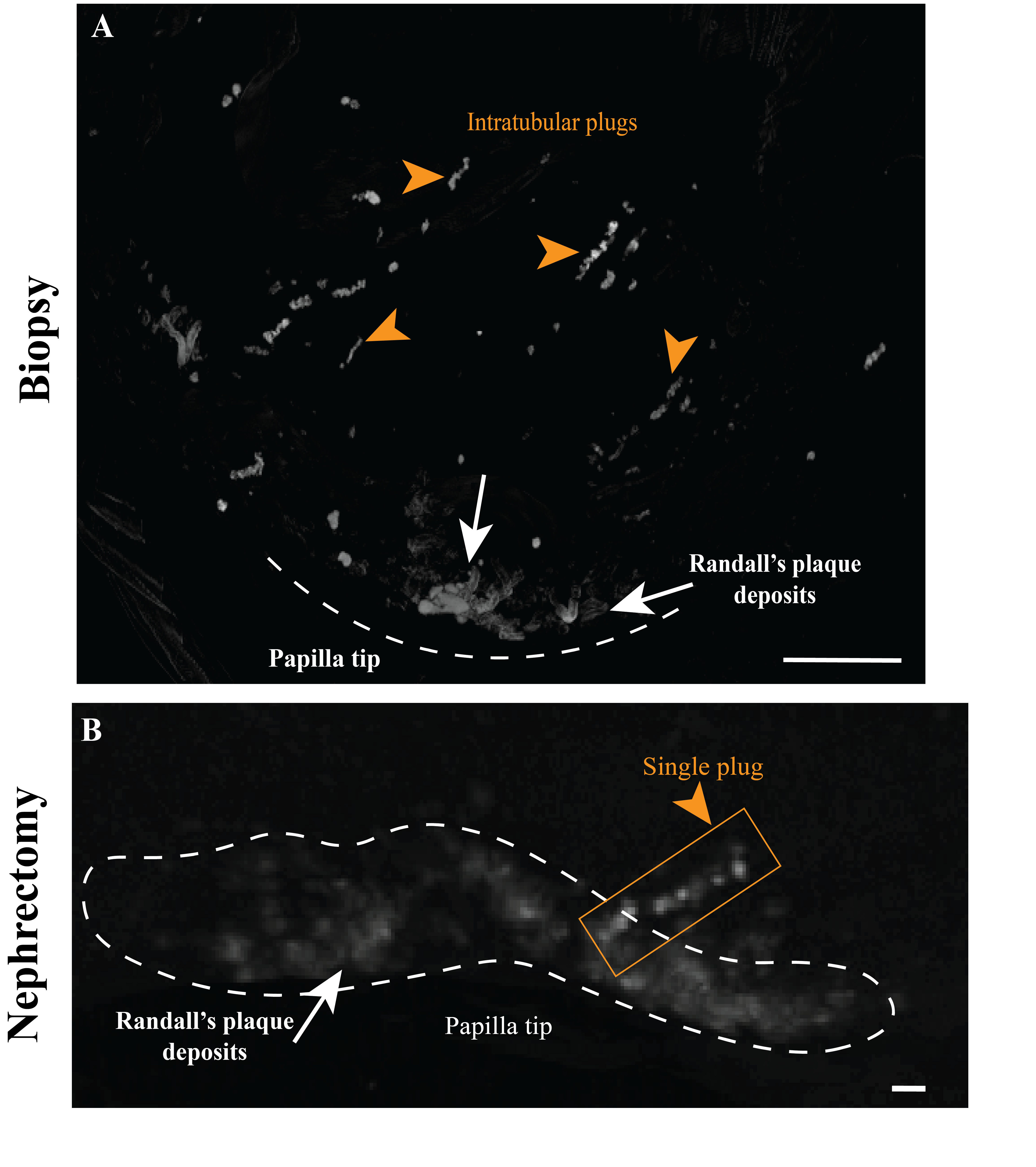
Introduction: Interstitial mineralization in the renal papilla tip is associated with retention and early kidney stone formation. A recent hypothesis proposes that upstream mineral plugging of renal tubules initiates downstream calcifications that lead to formation of Randall’s plaques (RP) at the papilla tip. The objective of the present study was to test this hypothesis by assessing the correlation between upstream mineral plugs and downstream RP formations at the papilla tip. Methods: Papilla specimens (22 in total, including 16 biopsies from 11 patients and 6 papillae from 5 nephrectomy samples) were scanned by micro computed tomography (CT) and measurements were collected by two investigators blinded to the number of plugs and to the presence of RP. Results: Of the 22 papillae, 3 nephrectomy specimens showed RP by micro CT along with 6.0±5.6 plugs (range, 1 to 12 plugs), with an average plug distance of 1.8±0.89 mm from the papilla tip. The remaining 3 nephrectomy specimens did not contain RP but still displayed 6.7±2.25 plugs (range, 5 to 9 plugs) and an average distance of 2.4±1.7 mm from the papilla tip. Ten (10) biopsy specimens showed evidence for RP along with 12.3±15.1 plugs (range, 1 to 51 plugs) and an average plug distance of 1.0±0.35 mm from the papilla tip, while 6 biopsy specimens without RP displayed 7.8±2.9 plugs with an average distance of 1.1±0.33 mm from the papilla tip. Our analyses revealed that neither the number of intratubular plugs nor the distances from the papilla tip to the plugs differed by the presence of RP at the papilla tip (p=0.72 and p=0.34, respectively). Conclusions: Our results suggest that RP is not correlated with upstream mineral formation. Further investigation on the development of RP using multi-omics and high-resolution imaging technologies may help to elucidate essential cellular and molecular effectors that drive the formation of RP in patients with stone disease. SOURCE OF Funding: Funded by: NIH P01 DK056788