
Poster Session C
Fibrosing rheumatic diseases (scleroderma, MCTD, IgG4-related disease, scleroderma mimics)
Sasha Shenk, BS
Tufts University
Boston, MA, United States
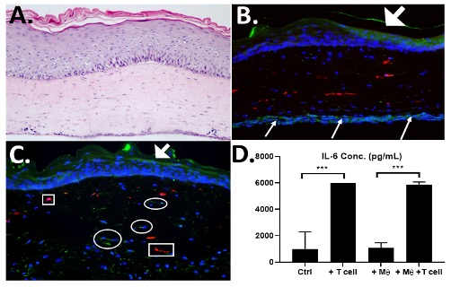 Figure 1. T cells in skin equivalents are functional- A) H&E image of 3D skin containing patient-derived fibroblasts, Macrophages and T cells. B) IHC staining for CD206-Macrophages (red) CD4-T cells (green) on bottom of tissue that migrated into dermis in 4 days (C). T cell function was shown by elevation of IL-6 secretion in T cell-containing tissues when compared to control or macrophage-only containing tissues (D).
 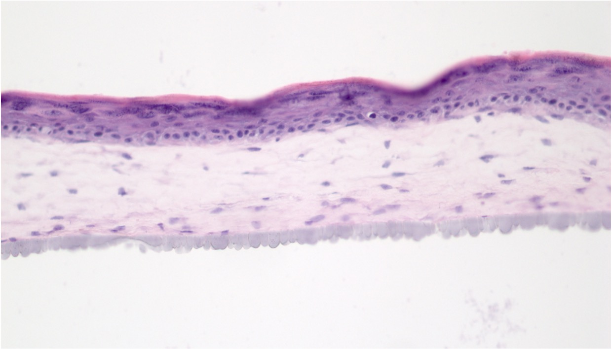 Figure 2. Fabrication of fully autologous human tissues with SSc cells- HSE skin-like tissues were constructed with patient-matched, SSc-derived keratinocytes (SK15.2) and fibroblasts (SF15.2). Tissues were grown for 2 weeks revealing well-differentiated epithelium and well-structured dermis.