
Poster Session B
Rheumatoid arthritis (RA)
Sonia Presti, BS
Brigham and Women's Hospital
Boston, MA, United States
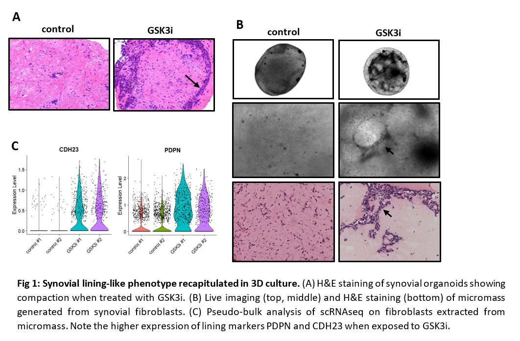 Fig 1: Synovial lining-like phenotype recapitulated in 3D culture. (A) H&E staining of synovial organoids showing compaction when treated with GSK3i. (B) Live imaging (top, middle) and H&E staining (bottom) of micromass generated from synovial fibroblasts. (C) Pseudo-bulk analysis of scRNAseq on fibroblasts extracted from micromass. Note the higher expression of lining markers PDPN and CDH23 when exposed to GSK3i.
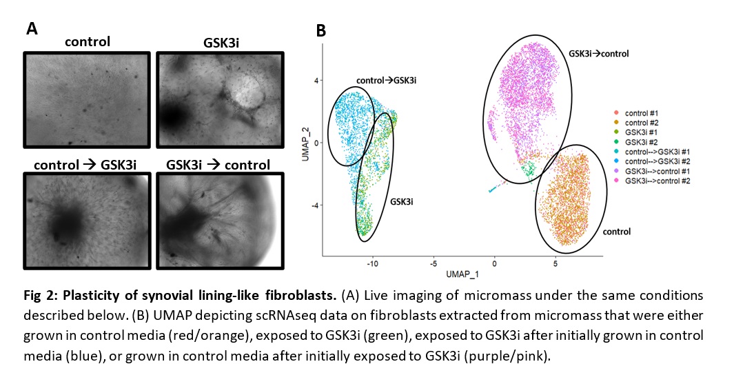 Fig 2: Plasticity of synovial lining-like fibroblasts. (A) Live imaging of micromass under the same conditions described below. (B) UMAP depicting scRNAseq data on fibroblasts extracted from micromass that were either grown in control media (red/orange), exposed to GSK3i (green), exposed to GSK3i after initially grown in control media (blue), or grown in control media after initially exposed to GSK3i (purple/pink).