
Poster Session A
Rheumatoid arthritis (RA)

Sahar Lotfi-Emran, MD, PhD
University of Minnesota
Minneapolis, MN, United States
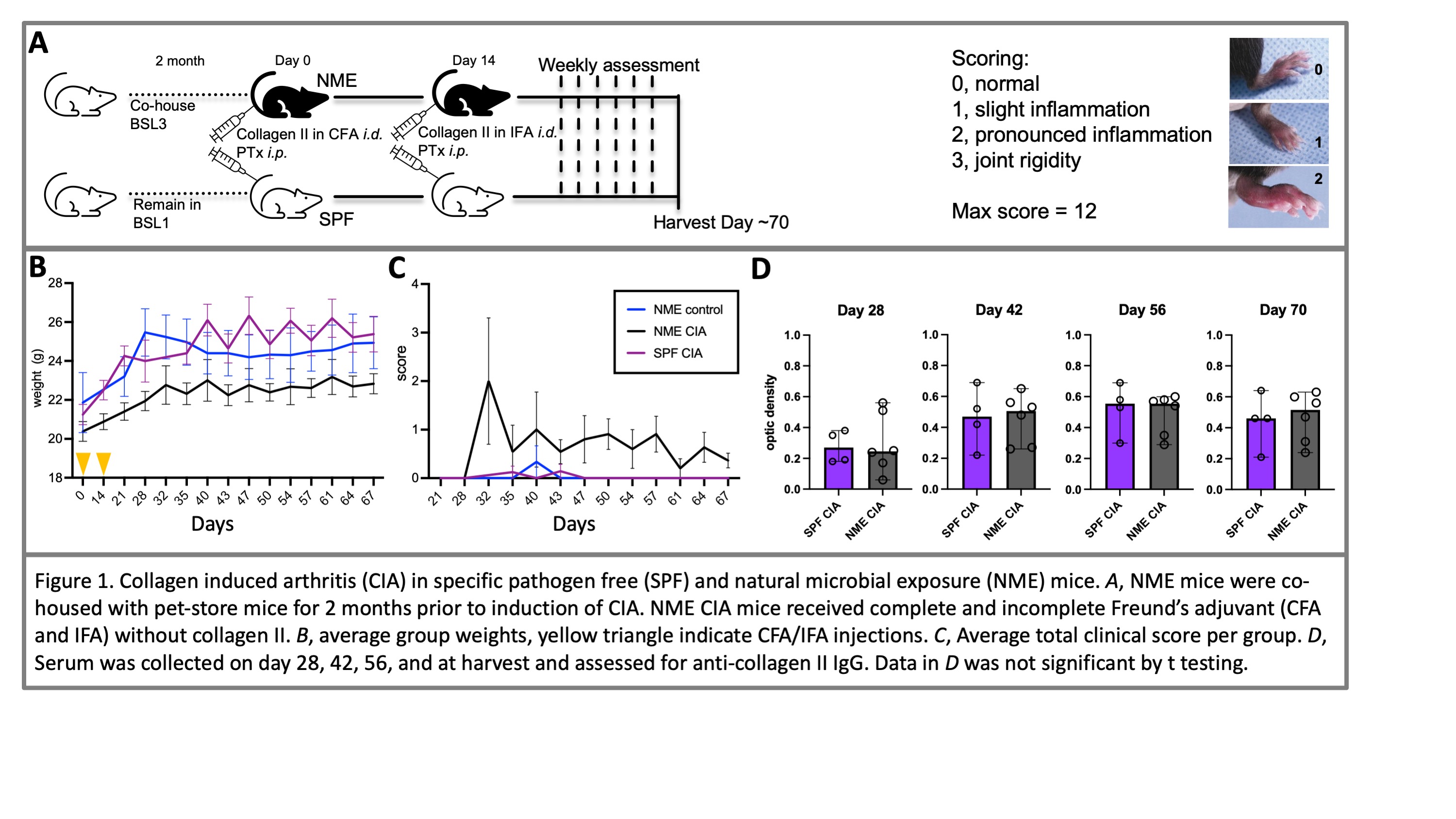 Figure 1. Collagen induced arthritis (CIA) in specific pathogen free (SPF) and natural microbial exposure (NME) mice. A, NME mice were co-housed with pet-store mice for 2 months prior to induction of CIA. NME CIA mice received complete and incomplete Freund’s adjuvant (CFA and IFA) without collagen II. B, average group weights, yellow triangle indicate CFA/IFA injections. C, Average total clinical score per group. D, Serum was collected on day 28, 42, 56, and at harvest and assessed for anti-collagen II IgG. Data in D was not significant by t testing.
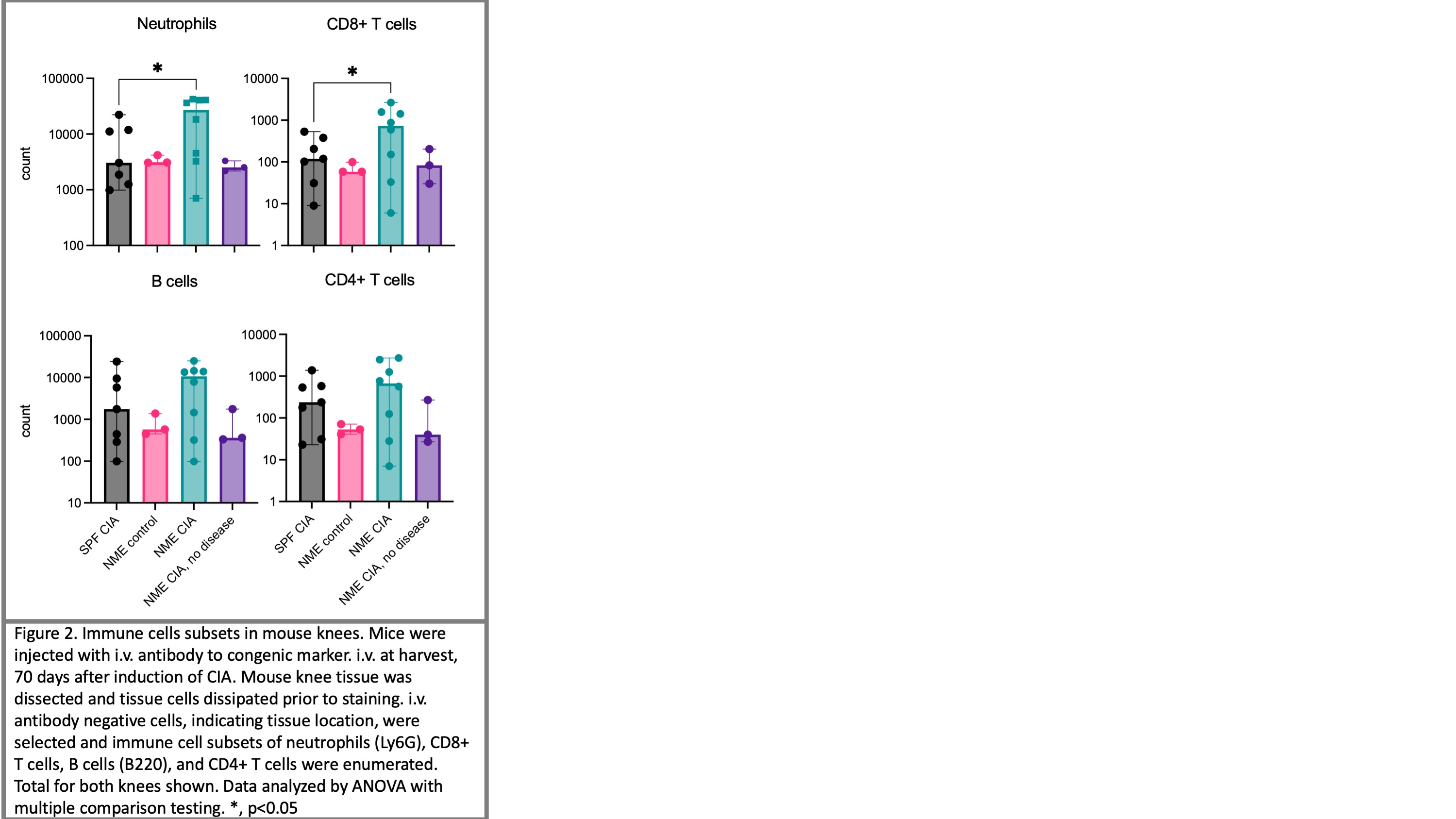 Figure 2. Immune cells subsets in mouse knees. Mice were injected with i.v. antibody to congenic marker. i.v. at harvest, 70 days after induction of CIA. Mouse knee tissue was dissected and tissue cells dissipated prior to staining. i.v. antibody negative cells, indicating tissue location, were selected and immune cell subsets of neutrophils (Ly6G), CD8+ T cells, B cells (B220), and CD4+ T cells were enumerated. Total for both knees shown. Data analyzed by ANOVA with multiple comparison testing. *, p < 0.05
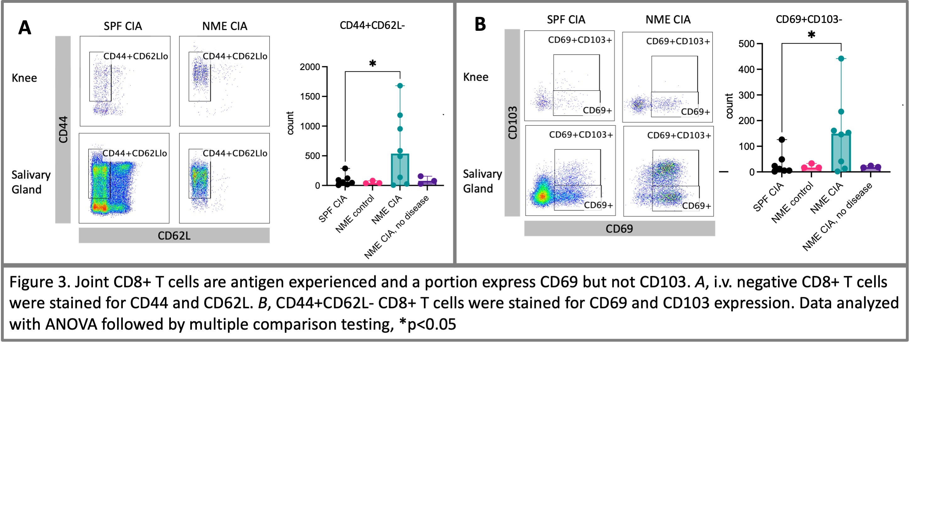 Figure 3. Joint CD8+ T cells are antigen experienced and a portion express CD69 but not CD103. A, i.v. negative CD8+ T cells were stained for CD44 and CD62L. B, CD44+CD62L- CD8+ T cells were stained for CD69 and CD103 expression. Data analyzed with ANOVA followed by multiple comparison testing, *p < 0.05