
Poster Session C
Fibrosing rheumatic diseases (scleroderma, MCTD, IgG4-related disease, scleroderma mimics)
Amela Hukara, MSc
University Hospital Zurich, University of Zurich
Schlieren, Switzerland
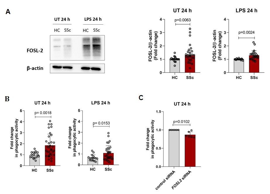 Figure 1: FOSL-2 protein levels and phagocytosis in HC and SSc hMDM. A) Representative Western Blot image and fold changes of FOSL-2 protein levels for unstimulated (UT) and LPS stimulated hMDM, n= HC (11-18), SSc (18-25). B) Fold changes of phagocytic activity are shown for unstimulated (UT) and LPS stimulated hMDM, n= HC (12-16), SSc (29-34). C) Fold changes of phagocytic activity are shown for control siRNA and FOSL2 siRNA silenced unstimulated (UT) hMDM, n= (6).