
Poster Session B
Rheumatoid arthritis (RA)
.jpg "Nozima Aripova, BS photo")
Nozima Aripova, BS
University of Nebraska Medical Center
Omaha, NE, United States
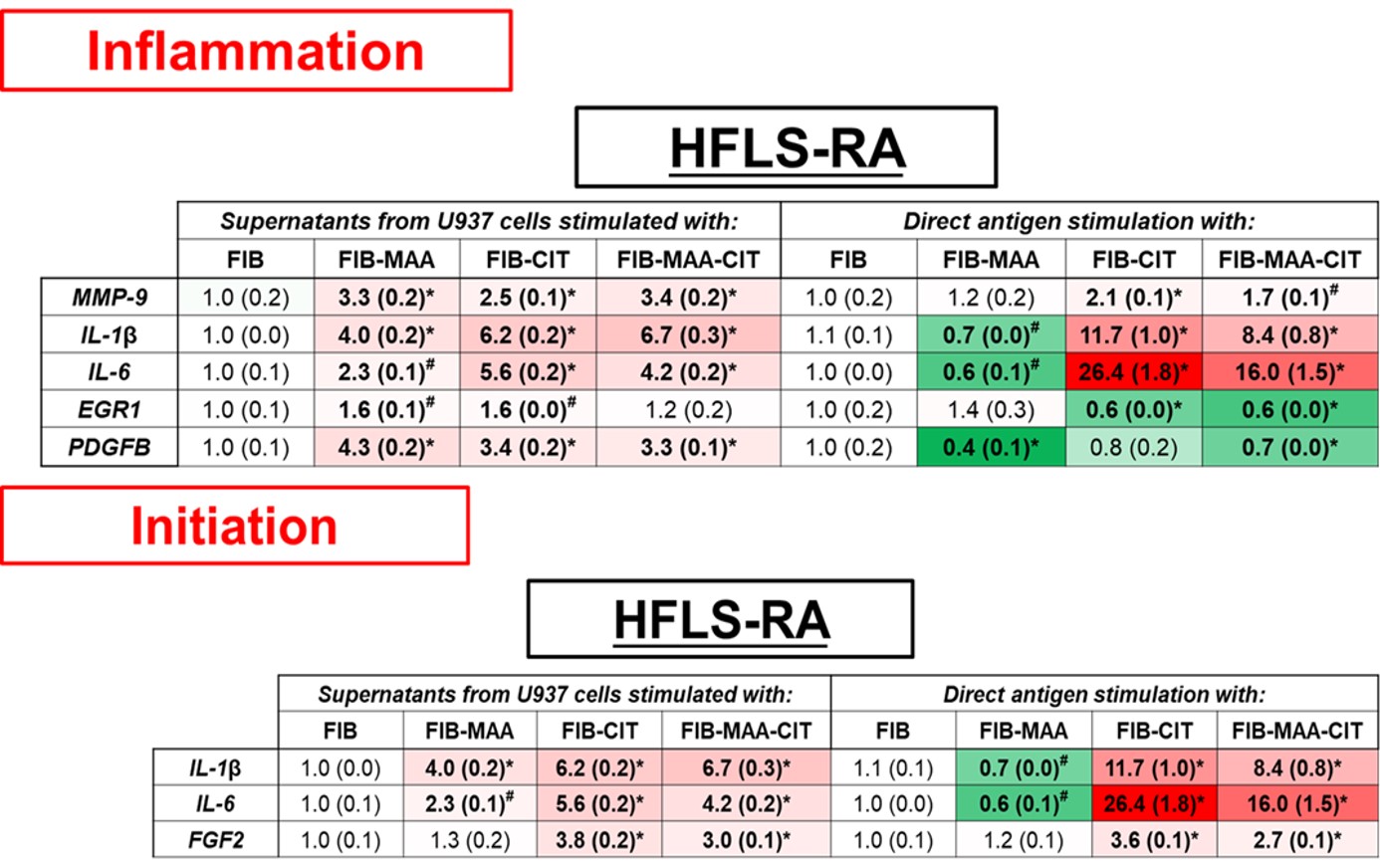 Figure 1. PCR for mRNA levels of inflammation and initiation fibrosis markers from stimulated HFLS-RA cells. HFLS-RA cells were stimulated with either supernatants from modified antigens treated U937 cells or with directly modified antigens. The data is represented as mean (standard deviation) of relative quantity (Rq) of fibrosis markers. All the values are compared to native protein. #p < 0.05, *p < 0.001, n=3.
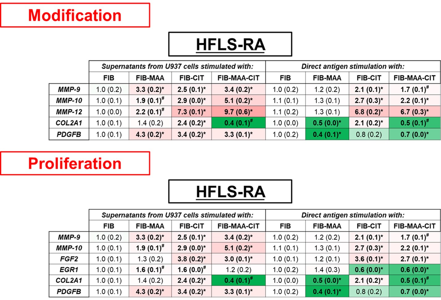 Figure 2. PCR for mRNA levels of modification and proliferation fibrosis markers from stimulated HFLS-RA cells. HFLS-RA cells were stimulated with either supernatants from modified antigens treated U937 cells or with directly modified antigens. The data is represented as mean (standard deviation) of relative quantity (Rq) of fibrosis markers. All the values are compared to native protein. #p < 0.05, *p < 0.001, n=3.