
Ignite Talk

Andrea Fava, MD
Johns Hopkins University
Baltimore, MD, United States
Disclosure: Disclosure(s): No financial relationships with ineligible companies to disclose
.jpg) Figure 1. Prevalence and concentrations of autoantibodies at lupus nephritis (LN) diagnosis according to LN classification. (A) Overall prevalence (any positive titer) of autoantibodies at the time of kidney biopsy in all patients with LN (class I-VI), any proliferative (class III, IV, III+V, or IV+V), and membranous (class V). (B-D) Autoantibody titers that significantly differ between patients with mesangial (class I), pure membranous (class V), pure proliferative (class III or IV), mixed (class III+IV or class IV+V), and advanced sclerosis (class VI) LN. Statistical significance was determined using Kruskal-Wallis test with Dunn’s multiple comparisons. *p < 0.05, **p < 0.01, ***p < 0.001, ****p < 0.0001
Figure 1. Prevalence and concentrations of autoantibodies at lupus nephritis (LN) diagnosis according to LN classification. (A) Overall prevalence (any positive titer) of autoantibodies at the time of kidney biopsy in all patients with LN (class I-VI), any proliferative (class III, IV, III+V, or IV+V), and membranous (class V). (B-D) Autoantibody titers that significantly differ between patients with mesangial (class I), pure membranous (class V), pure proliferative (class III or IV), mixed (class III+IV or class IV+V), and advanced sclerosis (class VI) LN. Statistical significance was determined using Kruskal-Wallis test with Dunn’s multiple comparisons. *p < 0.05, **p < 0.01, ***p < 0.001, ****p < 0.0001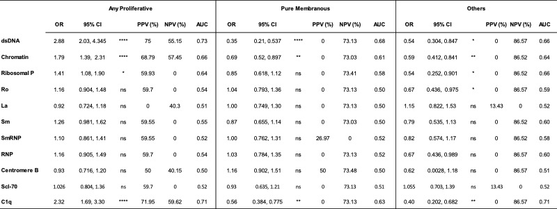 Table. Diagnostic implications of autoantibody titers at the time of kidney biopsy. For each autoantibody (rows), odds ratios (OR), positive and negative predictive values (PPV and NPV, respectively), and area under the curve (AUC) were calculated for each increase in titer by 1 standard deviation in patients with any proliferative (class III, IV, III+V or IV+V), pure membranous (class V), and other LN classes (class I, II, or VI)