
Ignite Talk
Guy Katz, MD
Massachusetts General Hospital
Boston, MA, United States
Disclosure: Disclosure(s): No financial relationships with ineligible companies to disclose
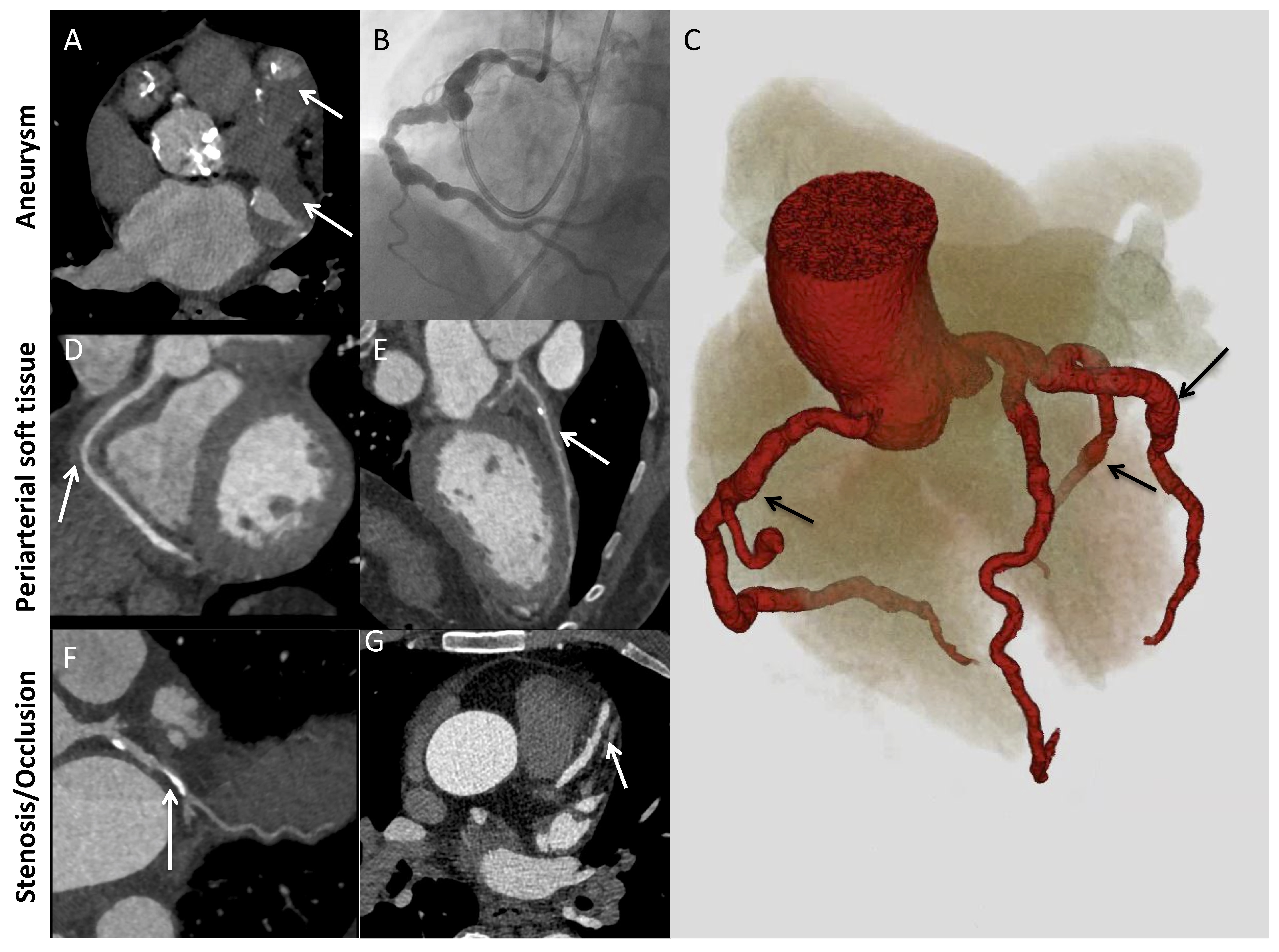 Figure 2. Major patterns of coronary artery involvement in IgG4-related disease. Images include axial CT (A), left heart catheterization (B), 3D volume-gated reconstruction of CTA (C), and curved planar reformatting of coronary CTA (D-G). Findings include fusiform aneurysmal dilatation (A-C) with mural thrombi (A, arrows), ectasia (C) periarterial soft tissue (D, E; arrows), and stenosis (F, G; arrows). Panel C demonstrates ectasia of the entire coronary tree with focal fusiform aneurysmal dilatations (arrows).
 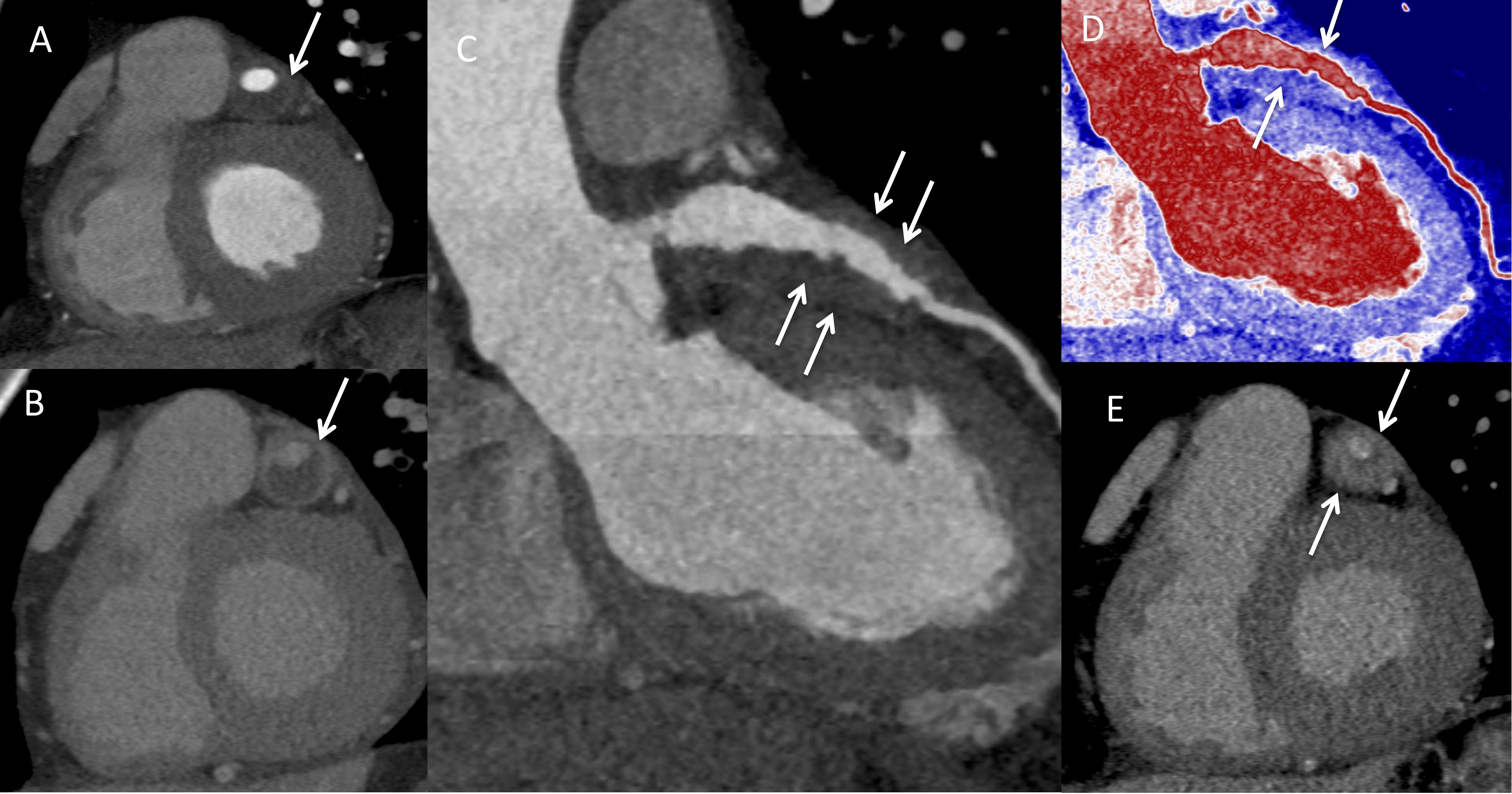 Figure 3. Arteritis and periarteritis. A) Coronary computed tomography (CTA) in the short-axis view demonstrating the left anterior descending artery (LAD) aneurysm (arrow). B) Delayed contrast-enhanced CT showing enhancement of the aneurysmal LAD wall consistent with arteritis (arrow). C) LAD curved planar reformatted (CPR) and D) Pseudo colorized image of the LAD CPR image showing the soft tissue thickening around the LAD (arrows). E) Delayed phase image in short axis showing enhancement of the soft tissue thickening consistent with periarteritis (arrows).