
Poster Session C
Systemic lupus erythematosus (SLE)

Erik Anderson, MD, PhD
Feinstein Institutes for Medical Research
New York, NY, United States
.jpg) Table: Demographics and characteristics of SLE subjects and healthy controls (HC) who underwent FDG-PET and DTI. Comparisons are between the 14 SLE subjects who underwent FDG-PET imaging (13 of these subjects also underwent DTI imaging) and 5 HC. All data is reported either as a mean (or median where indicated) +/- standard deviation (or interquartile range), or as a frequency (%). All data refers to that which was collected at the time of evaluation.
Table: Demographics and characteristics of SLE subjects and healthy controls (HC) who underwent FDG-PET and DTI. Comparisons are between the 14 SLE subjects who underwent FDG-PET imaging (13 of these subjects also underwent DTI imaging) and 5 HC. All data is reported either as a mean (or median where indicated) +/- standard deviation (or interquartile range), or as a frequency (%). All data refers to that which was collected at the time of evaluation.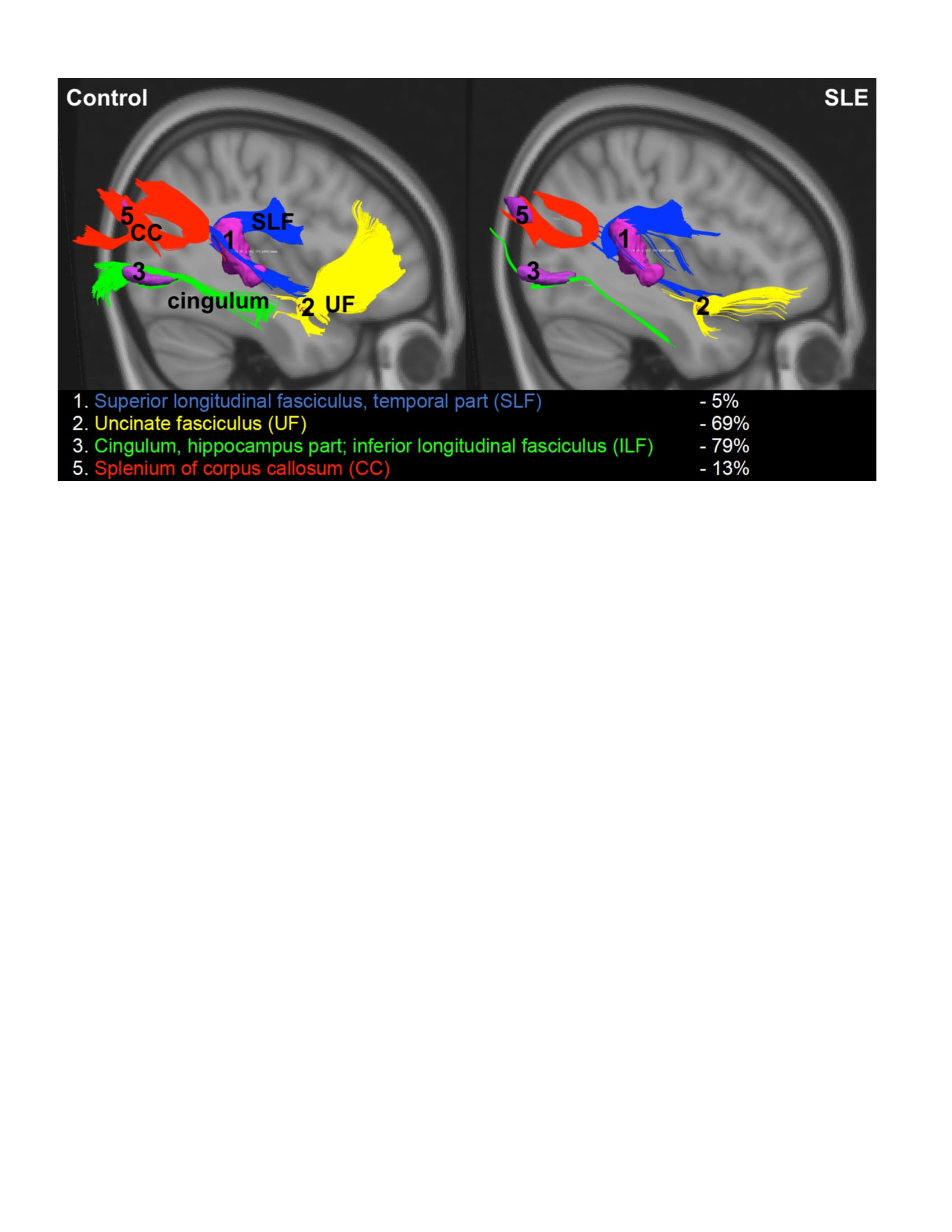 Figure: Group Tractography Displays Abnormal White Matter Pathways in SLE. SLE subjects demonstrated decreased fractional anisotropy (FA) in 4 of 5 previously identified abnormal FA clusters in SLE.[3] The images shown represent reconstructed white matter tracts between clusters of decreased white matter microstructural integrity (decreased FA). The percent reduction in white matter tract numbers in SLE versus HC is displayed.