
Poster Session A
Osteoarthritis (OA) and related disorders
Sung Yeon Kim, BS
University of Pennsylvania
Philadelphia, PA, United States
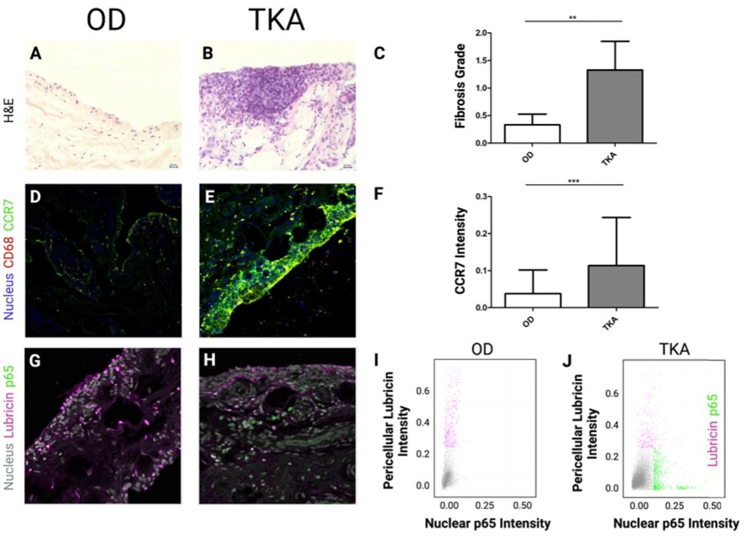 Figure 1: Representative H&E histology of human synovium from an (A) OD and (B) TKA patient and their (C) fibrosis scores. (D) OD and (E) TKA synovium stained for CD68, DAPI, and CCR7. (F) Quantification of CCR7 fluorescent intensity. (G) OD and (H) TKA synoviums stained for DAPI, PRG4 and p65. Single-cell analysis of lubricin vs. nuclear p65 intensity in (I) OD and (J) TKA synovium. **P < 0.01 and ***P < 0.001
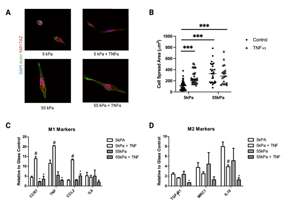 Figure 2: (A) Representative images of THP-1 macrophages cultured on 5 or 55 kPa polyacrylamide gels in the presence or absence of TNF-ɑ and their corresponding (B) cell spread area. Expression of (C) pro-inflammatory M1 and (D) pro-regenerative M2 markers. *P < 0.05 when comparing substrates with the same soluble condition and #P < 0.05 when comparing TNF-ɑ to no TNF-ɑ with the same substrate condition.
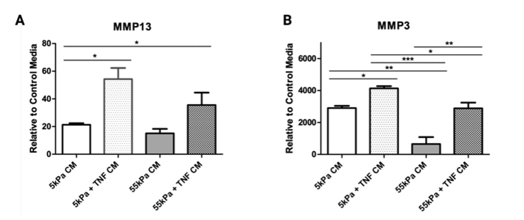 Figure 3: Expression of (A) MMP13 and (B) MMP3 in hMSCs cultured in THP-1 conditioned media on 5 and 55 kPa with and without TNF stimulation. *P < 0.05, **P < 0.01 and ***P < 0.001