
Abstract Session
Rheumatoid arthritis (RA)
Roopa Madhu, PhD
Brigham and Women's Hospital
Brookline, MA, United States
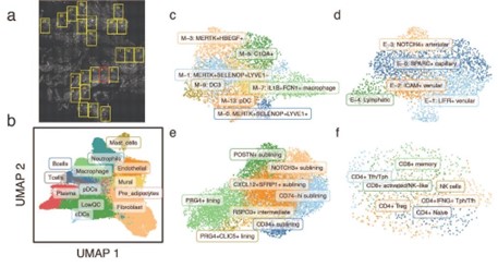 Identification of cell states in RA synovia using single-molecule spatial transcriptomics. (a) Highlighted FOVs were profiled with CosMx. (b) UMAP projection shows the location of individual cells in low dimensional space, colored by their inferred major cell type. Fine-grained clustering results of (c) myeloid cells, (d) endothelial cells, (e) fibroblasts, and (f) T cells are shown in fine-grained UMAP plots.
 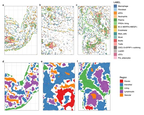 Spatial reconstruction of cellular networks in RA synovia. (a-c) Three representative fields of view images of CosMx RA synovial dataset shows anatomical distribution of major cell types and selected cell subtypes. (d-f) In the same images, spatially localized cells were grouped into five distinct anatomical regions