
Poster Session C
Imaging
Chirag Rajkumar Kopp, MD, MBBS, DM
Post Graduate Institute of Medical Education and Research
Lucknow, Uttar Pradesh, India
.jpg)
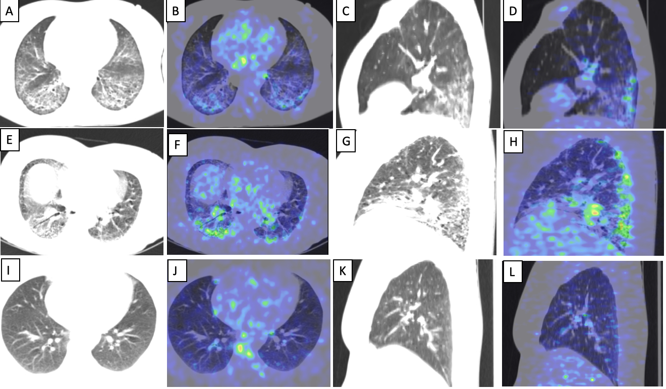 In patient with stable disease, transaxial CT images (A) showing reticular opacities with subpleural sparing and areas of architectural distortion with increased tracer uptake on the fused PET/CT images (B). Reformatted sagittal CT (C) and fused PET/CT (D) images showing the changes confined predominantly to the basal zones with relative sparing of the upper lobes. In patient with active disease, (E) showing reticular opacities and areas of patchy consolidation and ground glassing with increased tracer uptake on the fused PET/CT images (F). Reformatted sagittal CT (G) and fused PET/CT (H) images showing similar changes predominantly in the posterior lung fields with focal areas of architectural distortion
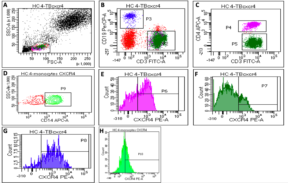 Representative flow diagrams showing gating strategy for CXCR4 expression: a) Gating of lymphocytes on FSC and SSC dot plot and monocytes were gated on cluster present between lymphocytes and granulocytes , b) showing CD3+ T cells as P2, CD19+ B cells as P3, c) showing CD4+ T cells as P4 and CD4- CD3+ (CD8 T cells) as P5 d) Showing CD14+ monocytes as P9, (e,f,g,h) showing CXCR4 expression on CD4+ T cells as p6, CD8+ T cells as p7, CD19+ B cells as p8 and CD14+ monocytes as p 10, respectively.