
Poster Session A
Osteoarthritis (OA) and related disorders
Meaghan Holub, BS
Central Michigan University College of Medicine
Mount Pleasant, MI, United States
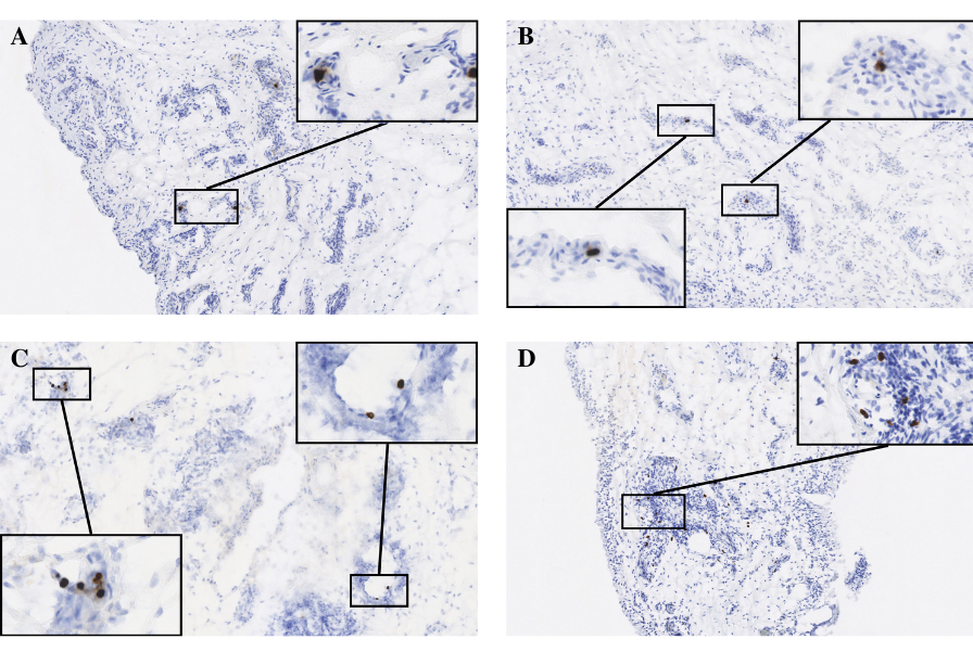 Figure 1: Synovial tissue with abundance of cells positive for bacterial PG (brown) per 10mm2 area was ranked according to a 4-point scale (0=none, 1=1-9 PG foci, 2=10-19 foci, 3=20-29 foci, 4=30+ foci). (A) Positive control – synovial tissue sample obtained from periprosthetic joint infection patient (B) Score 2 – PG positive cells surrounded by foci of inflammatory infiltrate (blue) (C) Score 3 – PG positive cells (brown) show selective accumulation surrounding various points of vasculature in synovial tissue sample (D) Score 4 – abundant accumulation of PG positive cells (brown) in tissue segment containing high-density inflammatory infiltrate (blue) and blood vessels.