
.png)
Trauma

Kaitlin Fusco, DDS
Resident
UCLA
Los Angeles, California, United States
Chi-Hong Tseng, Ph.D, MS
UCLA, Division of General Internal Medicine and Health Services Research
Itzel Rodriguez, BDS
UCLA Pediatric Dentistry
Daniela R. Silva, DDS, MS
Program Director
UCLA
Los Angeles, California, United States
Purpose: The purpose of this study is to identify the radiographic frequency of traumatic dental injuries in the primary dentition occurring in patients five and younger, as well as any correlation with dental trauma to gender or medical history. In addition, a secondary outcome is to analyze parental awareness of dental trauma in the primary dentition, by recording if the radiograph was taken at an emergency or at a recall visit.
Methods: This retrospective study was conducted by analyzing digital maxillary periapical radiographs of patients ages 5 years 11 months and below at time of radiograph between 2012-2020. Electronic health records were reviewed to collect information regarding age, gender, and medical history. Dental trauma to primary incisors were noted with radiographic signs as (1) external inflammatory root resorption (2) root fracture (3) arrested dentin deposition (4) tube like mineralization (5) pulp canal obliteration (6) internal root resorption (7) periapical radiolucency. If a patient presented with dental trauma, the SOAP note at the date of the radiograph was evaluated for the type of visit. Patients with radiographic signs of caries, previous restorations, or pulp treatment were excluded from this study. Statistical data analysis will be performed using Fisher’s exact test with significance level set at P value < .05. Correlation analysis will be performed to identify primary tooth trauma with patient gender and medical history.
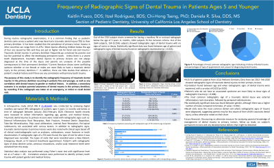
Results: Out of the 2703 subject charts screened for having a maxillary PA or occlusal radiograph below the age of 5 years 11 months, 1664 subjects fit the inclusion criteria. Out of the 1664 subjects, 154 (9.25%) subjects showed radiographic signs of dental trauma with no signs of caries or decay. Statistically significant data was found between age of patient and radiographic signs of dental trauma found on radiographic examination (p = 0.022).
Conclusion: Correlation was found between age and time of radiographic examination showing signs of dental trauma, with a p-value of 0.022 (p < 0.05). Patient’s who do not have an associated syndrome are more likely to show signs of radiographic trauma (p = 0.008). The most common radiographic sign of a traumatic dental injury was external inflammatory root resorption, followed by periapical radiolucencies. No statistically significant data was found between gender, although there was a higher number of males compared to females. (P value = 0.061). Recall visits were the most common dental visit where radiographic signs of trauma were diagnosed, suggesting parents may be less aware of their child’s traumatic dental injury, unless otherwise noted on their chart.
Future Research: Discovering an alternate measure for analyzing parental knowledge of management of dental trauma to primary incisors, follow up study on subject’s permanent incisors and any developmental defects that might be detected.