
.png)
Oral Pathology

Alaa Khoja, BDS
Pediatric dental resident
Boston University, Boston, MA
Boston University
Boston, Massachusetts, United States
Japriyaa R. Shanmugham, BDS, MPH, DrPH
Director of Research & Clinical Assistant Professor
Boston University Goldman School of Dental Medicine
Boston, Massachusetts, United States
Keri Discepolo, DDS, MPH
Post Graduate Program Director of Pediatric Dentistry
Boston University Goldman School of Dental Medicine
Boston, Massachusetts, United States
Introduction: Benign Fibrous Histiocytoma (BFH) is a soft tissue neoplasm, which mostly affects the skin of extremities. It rarely occurs in bones, and is mainly reported in femur, tibia, and pelvic bone. The occurrence in jawbones is very rare and may appear like odontogenic and non-odontogenic lesions of the maxilla and mandible.
Patient information/background: This report discusses a 13-year-old male patient who was first referred to BMC OS department for evaluation of a mandibular lesion after being incidentally noticed by his general dentist on the radiograph. Patient did not report any pain associated with the mandibular lesion. The patient did not have any pertinent past medical, family, or surgical history and there is no history of use of medications. The patient also did not report any drug allergies.
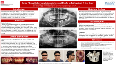
Clinical examination: Intra-oral exam revealed no buccal or lingual expansion, and missing mandibular anterior teeth due to history of extractions. The remaining dentition are not mobile. The lesion was asymptomatic, hard in consistency, and covered by normal-appearing oral mucosa. Panoramic radiograph and computed tomography showed a unilocular, well defined radiolucent, osteolytic lesion in the anterior mandibular region, crossing the midline, causing some root resorption, displacement, and cortical bone erosion. Incisional biopsy was performed under general anesthesia, and histopathologic analysis revealed BFH of the bone.